πŸ”’ Open the interactive required-margin demo →
Virtual Human Brain Β· Neurosurgical Planning & Robotics

The safest path to the target,
computed β€” not guessed.

Sulcora turns a patient's MRI into an AI-guided planning environment: name a target and it recommends the safest entry, replaces the fixed 2–3 mm margin with a computed, per-structure required standoff that accounts for predicted brain shift and the robot's own targeting error, simulates the insertion, and serves as a validation bench for neurosurgical robotics.

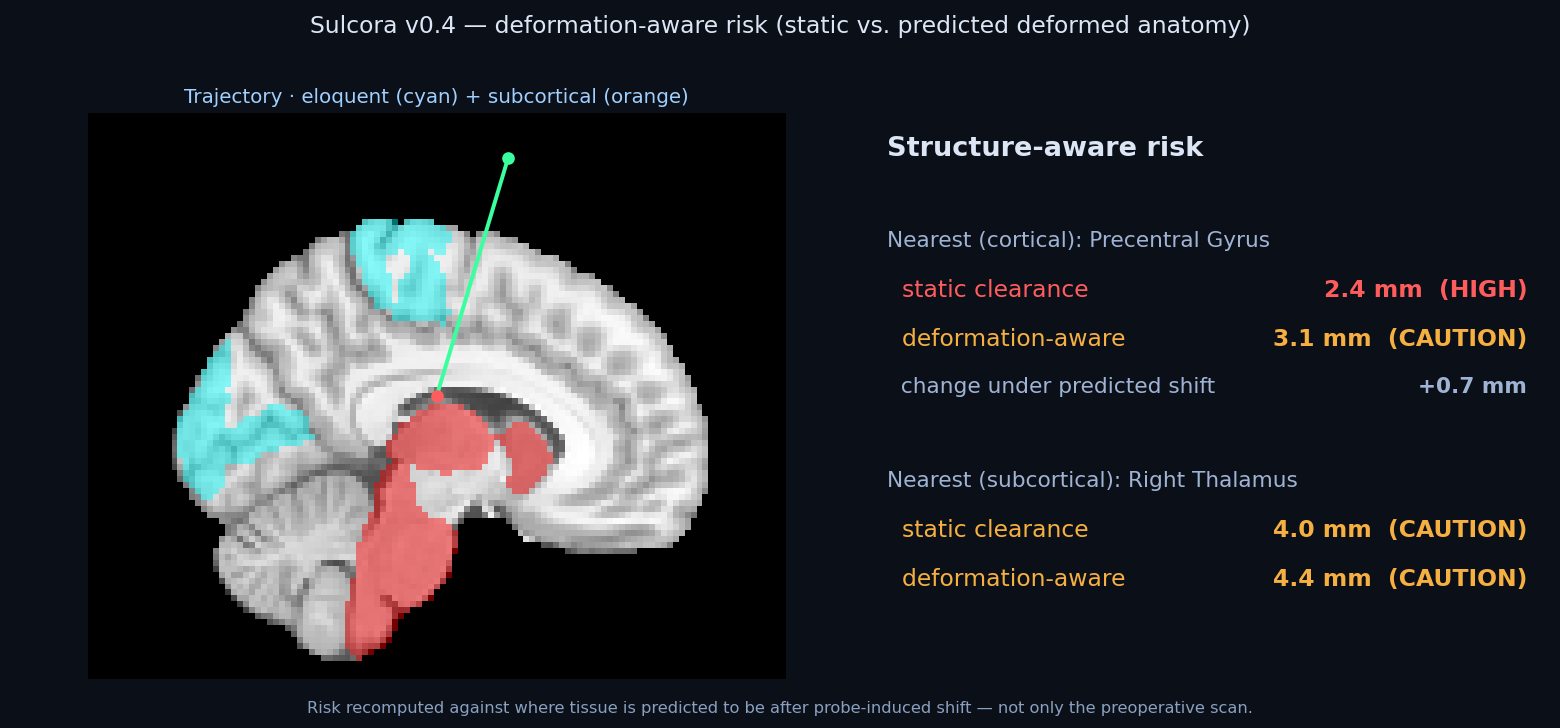
Sulcora deformation-aware risk on a real MRI
Live output: automated trajectory with structure-aware risk recomputed against predicted tissue deformation β€” on a real human MRI and the Harvard-Oxford anatomical atlas.
30
critical structures (incl. tracts)
~2,600
entry corridors searched / target
Real MRI
MNI152 + Harvard-Oxford atlas
20+
integrated capabilities
The problem

Planning a path into the brain is unforgiving β€” and today's tools plan on a brain that no longer exists.

Reaching a deep target means threading a straight line past eloquent cortex, vessels, and tracts. Conventional navigation plans on the preoperative scan, but the brain shifts the moment an instrument enters β€” so the safety margin you planned isn't the one you get. And the field is racing toward surgical robots with no high-fidelity virtual brain to develop and validate them against. Sulcora is built for both gaps.

Capabilities

A complete, working planning toolkit

Everything below runs today on real imaging β€” not mock-ups. Try it in the live planner.

POINT OF NOVELTY

Computed required standoff

Instead of one fixed 2–3 mm margin for every structure, Sulcora computes a per-structure, anisotropic required standoff β€” inverting a contact-probability model that fuses the robot's targeting-error distribution with the signed, toward-corridor component of predicted tissue shift. A structure predicted to deform toward the corridor demands more margin; one deforming away, less. It now drives the optimizer's ranking, not just a demo.

NEW

Uncertainty-aware margin

The margin comes with error bars: deformation-model uncertainty (from an ensemble of displacement fields) and CT↔MR registration error are propagated into the same probability inversion, in quadrature β€” so the standoff widens when the model is less certain, with a worst-case "stays safe even if the robot is 50% worse than spec" view instead of one over-confident number.

ROBOTICS

Vendor-neutral device qualification

Answers the question no planner asks: is THIS robot accurate enough for THIS case? Sulcora computes the maximum targeting error the corridor tolerates under predicted shift, passes/fails a specific device (e.g. "corridor needs ≀1.2 mm; a 0.9 mm robot PASSES"), and exports the keep-out geometry it must honor.

🧬

Multi-series fusion (CT↔MR)

Plan the corridor on the CT and pull eloquent cortex and tracts from a co-registered MR β€” or CTA vessels β€” into one self-consistent margin, via rigid mutual-information registration across modalities. The real multi-modal case, not one scan at a time.

βš–οΈ

Plan-vs-plan comparison

Load the corridor you drew and Sulcora shows where it agrees, where it recommends safer, and why β€” the binding structure and the headroom difference. It justifies against your plan instead of silently re-planning.

🎯

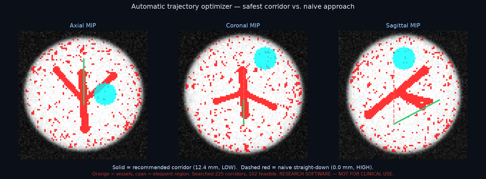
Automatic trajectory optimizer

Name a deep target β€” including real DBS targets (subthalamic nucleus, globus pallidus) β€” and Sulcora scores candidate entry corridors against clearance to vessels, eloquent cortex, and tracts at once β€” optionally against predicted deformation β€” recommending the safest path with ranked alternatives, under per-structure margin, angle, and depth limits.

πŸ”­

Probe-aligned views

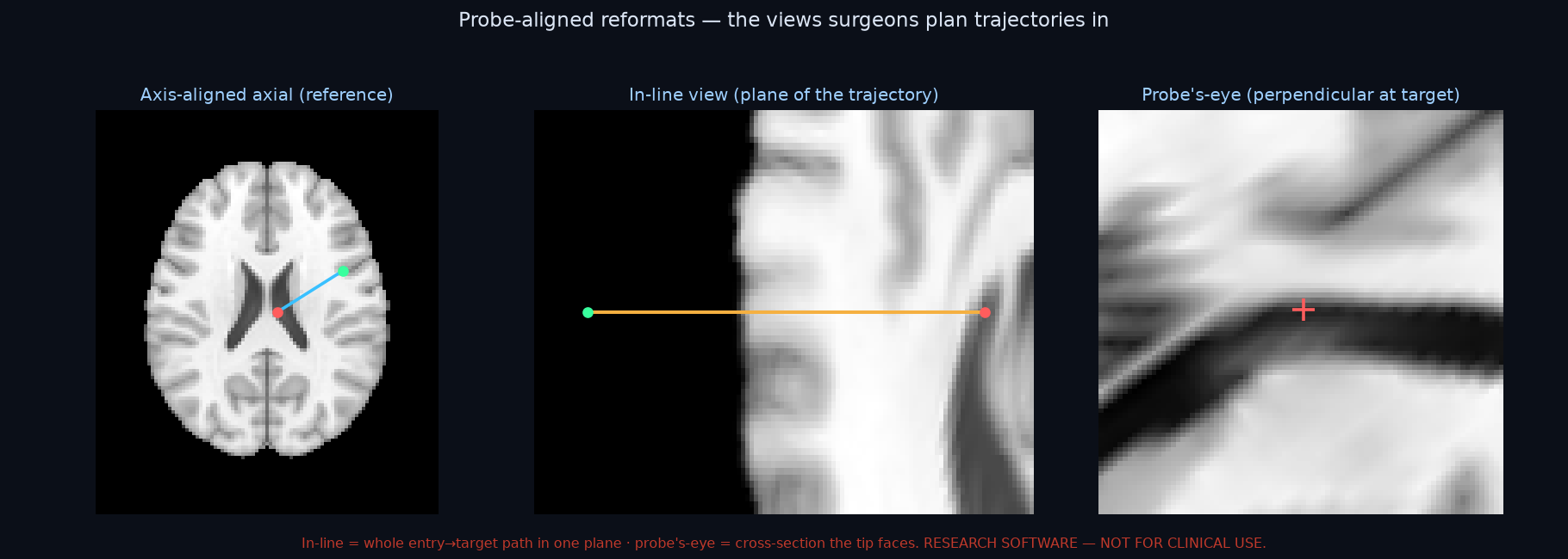
Beyond axial/coronal/sagittal — the in-line view (the whole entry→target path in one plane) and the probe's-eye view (the cross-section the advancing tip faces): the reformats surgeons actually plan trajectories in.

🧠

Structure-aware risk

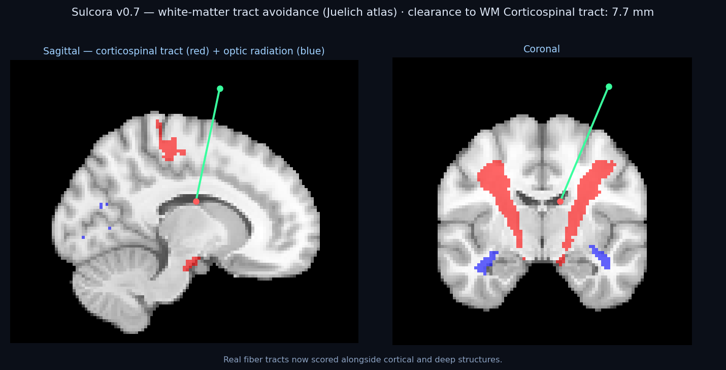
Real atlases: eloquent cortex (motor, speech, vision), deep structures (thalamus, hippocampus, brainstem), and white-matter tracts (corticospinal, optic radiation). Reports nearest structure, clearance, risk band.

🧩

Patient-specific segmentation

Optionally label eloquent cortex, ventricles, and deep structures directly from the patient's own T1 with a pretrained model (TotalSegmentator / SynthSeg) β€” registration-free, no atlas warp.

🩸

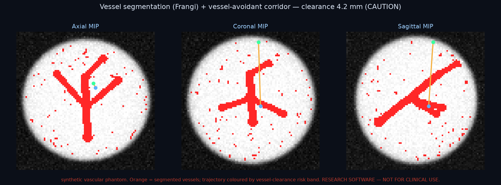
Vessel avoidance

Segment the vasculature from CTA / MR-angiography (multiscale vesselness) and report clearance to the vessel tree β€” including against the predicted deformed anatomy. Hitting a vessel is the dominant depth-electrode and biopsy risk.

DIFFERENTIATOR

Deformation-aware risk

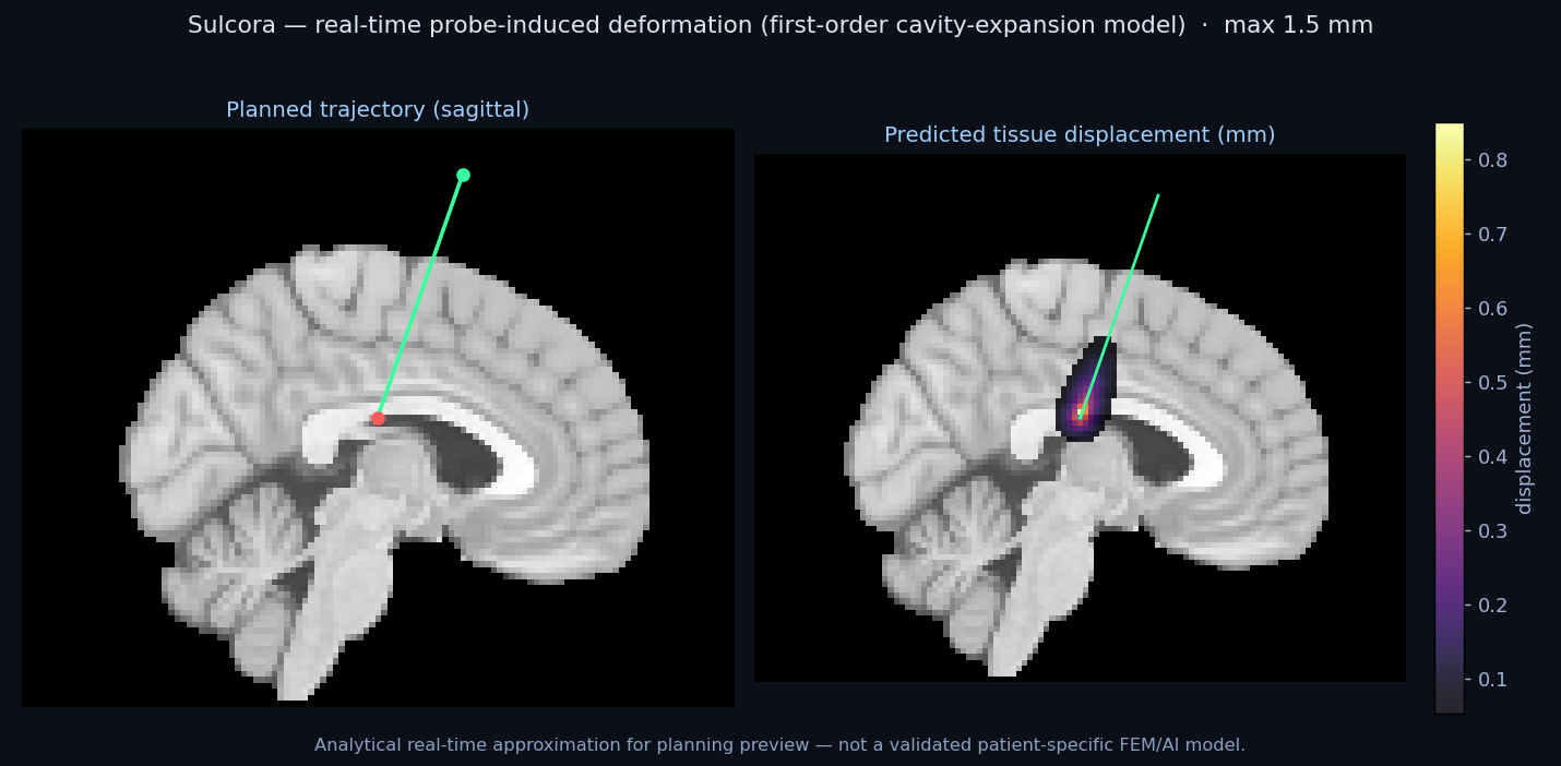
Risk recomputed against the brain's predicted deformed anatomy β€” the shift conventional static planning ignores.

🌊

Finite-element tissue deformation

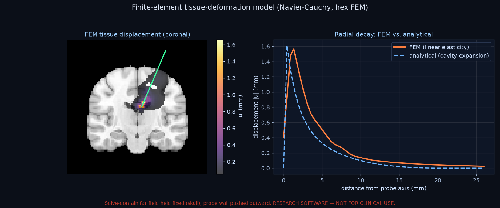
Predicts probe-induced tissue displacement by solving the elasticity equations on a finite-element mesh (Navier-Cauchy, hexahedral FEM) β€” upgrading the real-time analytical preview toward the validated model.

🎬

Insertion simulation

Advance the probe entry→target and watch clearance to critical structures at every depth, in all views.

ARPA-H TOPIC 7

Robotics-validation bench

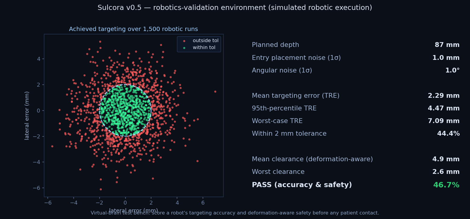
Score a simulated robot's targeting accuracy and deformation-aware safety over thousands of runs β€” before any patient contact.

⚿

Multi-trajectory planning

Plan multiple leads at once β€” bilateral DBS, multiple SEEG electrodes.

STROKE / ICH

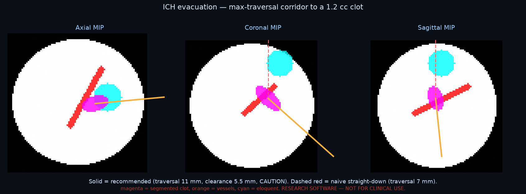
Haemorrhage (ICH) evacuation

Segment an acute clot from CT, find its long axis, and plan the corridor that maximises clot traversal while clearing vessels and eloquent cortex β€” the minimally invasive approach shown to improve outcomes in the ENRICH trial (NEJM 2024).

🩹

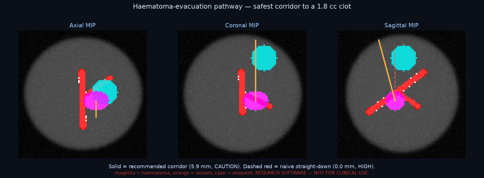
Lesion procedures

Beyond DBS/SEEG: target any segmented lesion by its centroid (volume in cc) for the safest corridor β€” biopsy, catheter/laser placement, or tumour access.

πŸ“„

Rich OR plan report

One-click PDF for the chart: per trajectory β€” tri-planar and probe-aligned reformats, a clearance-vs-depth profile, full geometry, and a per-structure clearance table (static and deformation-aware).

πŸ“

Validation & interop

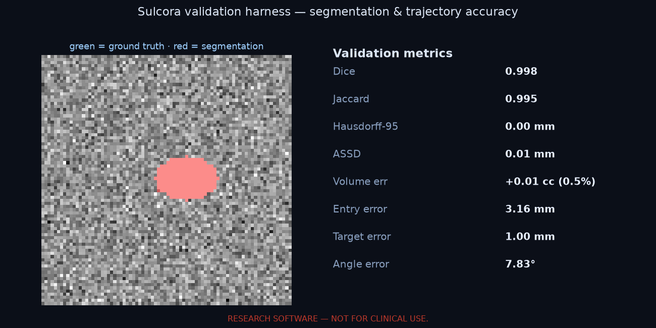
Quantitative accuracy metrics (Dice, Hausdorff-95, ASSD, volume & trajectory error) for evidence before clinical validation, and one-click export of the plan to 3D Slicer and a DICOM RT Structure Set for Brainlab / StealthStation.

🩻

Full CT support

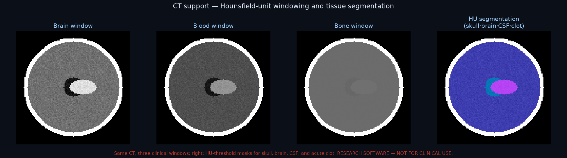
Head CT is the modality for haemorrhage and trauma β€” Sulcora loads it in Hounsfield Units with clinical window presets (brain / blood / bone / stroke) and HU-threshold tissue segmentation (skull, brain, CSF/ventricles, acute clot).

πŸ”’

Local & private

Runs on the surgeon's own machine β€” DICOM (from PACS) or NIfTI opened from disk. The patient image is processed on-device and never leaves it: no cloud upload, no PHI in transit.
Conditions & procedures

What Sulcora plans

One planning engine β€” the safest corridor to a target while avoiding vessels, eloquent cortex, and white-matter tracts, scored against the brain's predicted shift β€” generalises across cranial procedures:

Built

Intracerebral haemorrhage (ICH)

Stroke / haemorrhage clot evacuation: CT clot segmentation β†’ long-axis, max-traversal corridor avoiding vessels and eloquent cortex (ENRICH-supported).

Built

Deep brain stimulation (DBS)

Named deep targets (STN, GPi, …) β†’ safest entry with ranked alternatives.

Built

SEEG / epilepsy

Multi-electrode depth trajectories with vessel and structure avoidance.

Built

Biopsy & LITT

Lesion-centroid targeting for stereotactic biopsy and laser ablation corridors.

Built

Tumour / lesion access

Any segmented lesion (volume in cc) β†’ safest access corridor.

Built

Ventricular catheter (EVD / shunt)

Segment the ventricles, locate the frontal horn, and plan/score a catheter β€” cannulation, length, and clearance.

Built

Glioblastoma / glioma resection

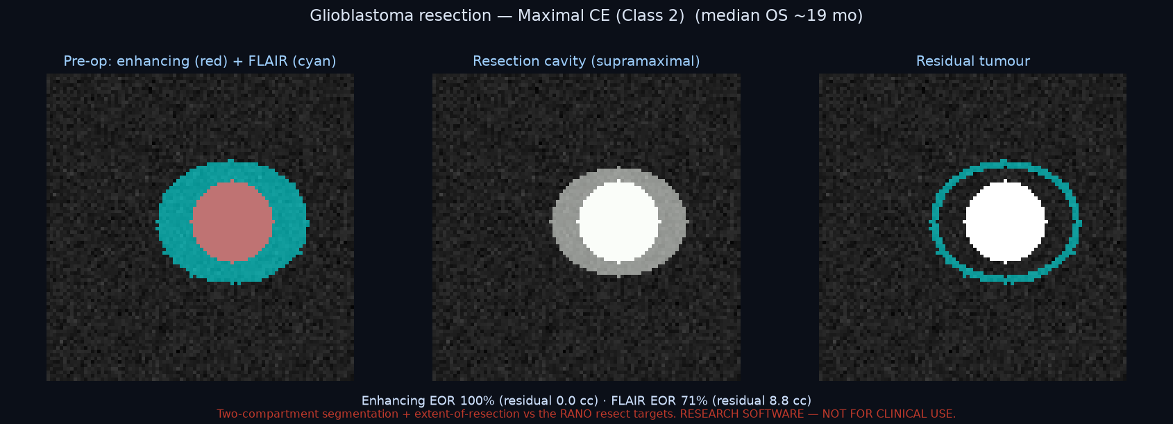
Two-compartment segmentation (enhancing + FLAIR infiltration) and extent-of-resection volumetrics with RANO resect grading. Awake-mapping integration in development.

Trajectory procedures (ICH, DBS, SEEG, biopsy, LITT, lesion access, EVD/shunt) run today in the toolkit; glioblastoma resection metrics (two-compartment segmentation, extent-of-resection, RANO grading) are in, with awake-mapping integration in development. The browser demo above shows deep-target planning on public data; the full per-procedure workflow (incl. CT/haematoma) runs in the local desktop app on your own scan.
Differentiation

Not a better viewer β€” a different question

Established navigation platforms are excellent at showing anatomy and tracking instruments. Sulcora adds the things they don't: planning against predicted intraoperative shift, and a virtual brain built for robotics. We integrate with the hardware surgeons already own β€” we don't replace it.

CapabilityConventional static planningSulcora
Multiplanar + 3D trajectory planningβœ“βœ“
Atlas-based eloquent-structure overlayβœ“βœ“
Automated "name a target β†’ safest entry"partial / manualβœ“ automated
Per-structure direction-dependent required margin (vs fixed 2–3 mm)fixed marginβœ“ computed
Uncertainty-quantified margin (deformation + registration error bars)β€”βœ“
Vendor-neutral device qualification (is the robot good enough for this case)β€”βœ“
Risk against predicted brain shiftβ€”βœ“
Vessel clearance vs predicted brain shiftβ€”βœ“
Finite-element tissue-deformation modelβ€”βœ“
Robotics development & validation benchβ€”βœ“
Insertion simulation with depth-wise clearancelimitedβœ“
Honest framing: Sulcora is differentiated on the highlighted axes; it is not yet a cleared clinical product, and conventional platforms lead on regulatory clearance, integration breadth, and validation today. Our differentiation is demonstrated in silico; retrospective real-data validation (below) shows calibrated safety and device qualification but no contact-rate superiority so far, so it is not yet a clinical claim.
Workflow

How a surgeon uses it

1

Name the target

Type a structure (e.g., Left Thalamus). Sulcora locates it on the patient's MRI.

2

Get the safest corridor

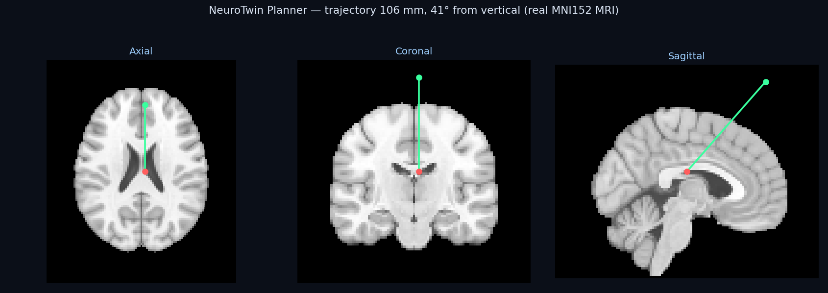
One click β€” the optimizer scores candidate entries against every critical structure at once and snaps to the recommended path, with ranked alternatives and the feasible-corridor count.

3

Verify & simulate

Linked crosshairs across axial/coronal/sagittal with a live risk readout β€” nearest structure, clearance, band, and the deformation-aware value β€” then simulate the insertion and export the plan report.

Validation & regulatory

An explicit path from research to cleared product

Sulcora is research software today. The route to clinical use is defined, staged, and the basis for collaboration with academic neurosurgery programs.

NOW βœ“

Working platform

Tested core, real-MRI planning, structure-aware + deformation-aware risk, finite-element deformation solver, patient-specific segmentation, robotics bench.

PHASE I

Bench + retrospective

Phantom accuracy (TRE), retrospective imaging vs expert plans, validated segmentation; peer-reviewed publication.

PHASE II

Cadaveric + robotics

Cadaveric validation, validated deformation model, hardware-in-the-loop robotics, QMS / IEC 62304.

CLEARANCE

FDA SaMD

510(k)/De Novo pathway, prospective clinical study, integration with navigation hardware.

Retrospective validation β€” results to date (in silico & public imaging). Stage 0 (seeded deformation-sensitive corridors) shows the deformation- and device-aware required standoff selecting lower-contact corridors than a fixed margin (~9% absolute reduction). On real public imaging β€” glioma MRI (UPENN-GBM), intracranial-haemorrhage CT (BHSD, expert-annotated clots), real stereo-EEG electrode trajectories from three institutions (610+ depth trajectories), and 100 real cerebral-vessel segmentations β€” the required standoff shows no mean contact-rate advantage over a fixed margin, and its recommended corridors agree with real implanted trajectories no better than chance (native-space paired p=0.80, unchanged by a population vessel atlas). The demonstrated value is the calibrated safety layer: after correcting the certificate’s contact model, its predicted contact-probability bound held as a proper upper bound for 100% of real trajectories across three SEEG institutions and 100 real vessel geometries, with vendor-neutral device qualification at 88–96%. On real MR angiography (99 TubeTK scans, 42 with expert vessel labels) and a second haemorrhage site (PhysioNet CT-ICH), the certificate stayed a valid upper bound in 610/612 device-error evaluations (99.7%); the software's own vesselness recovered 83% of large expert vessels but only 49% of fine ones, quantifying against expert ground truth why patient-specific angiography is still required. On the COSTA multi-center cerebrovascular dataset (six independent centers, 45 TOF-MRA with expert vessel masks) the certificate held in all 180 device-error evaluations, and the classical vesselness reached mean Dice 0.44 against the expert masks. And in the most stringent test yet, using patient-specific white-matter tracts from each subject's own diffusion MRI (33 subjects) in place of an atlas, the certificate stayed a valid upper bound across all 132 device-error evaluations even where contact reached 96%. We also validated the software's learned segmentation backend: trained on multi-center expert vessel masks and tested on held-out centers, it raised safety-relevant recall of fine (1–2 mm) vessels from 76% to 99% (strict voxel recall 27% to 93%), addressing the classical filter's fine-vessel gap. A real-data contact-rate benefit remains future work. These are feasibility and calibration results, not claims of clinical accuracy, safety, or efficacy.
Accuracy targets, study designs, and the regulatory pathway are detailed in the technical package (download below). Not a medical device; not for clinical, diagnostic, or treatment use.
Try it / get the package

See it work in your browser

Includes the application, tested core, sample real MRI, validation/clinical-study plan, and regulatory roadmap.