Everything below runs today on real imaging — not mock-ups. Try it in the live planner.
POINT OF NOVELTYComputed required standoff
Instead of one fixed 2–3 mm margin for every structure, Sulcora computes a per-structure, anisotropic required standoff — inverting a contact-probability model that fuses the robot's targeting-error distribution with the signed, toward-corridor component of predicted tissue shift. A structure predicted to deform toward the corridor demands more margin; one deforming away, less. It now drives the optimizer's ranking, not just a demo.
NEWUncertainty-aware margin
The margin comes with error bars: deformation-model uncertainty (from an ensemble of displacement fields) and CT↔MR registration error are propagated into the same probability inversion, in quadrature — so the standoff widens when the model is less certain, with a worst-case "stays safe even if the robot is 50% worse than spec" view instead of one over-confident number.
ROBOTICSVendor-neutral device qualification
Answers the question no planner asks: is THIS robot accurate enough for THIS case? Sulcora computes the maximum targeting error the corridor tolerates under predicted shift, passes/fails a specific device (e.g. "corridor needs ≤1.2 mm; a 0.9 mm robot PASSES"), and exports the keep-out geometry it must honor.
🧬
Multi-series fusion (CT↔MR)
Plan the corridor on the CT and pull eloquent cortex and tracts from a co-registered MR — or CTA vessels — into one self-consistent margin, via rigid mutual-information registration across modalities. The real multi-modal case, not one scan at a time.
⚖️
Plan-vs-plan comparison
Load the corridor you drew and Sulcora shows where it agrees, where it recommends safer, and why — the binding structure and the headroom difference. It justifies against your plan instead of silently re-planning.
🎯
Automatic trajectory optimizer
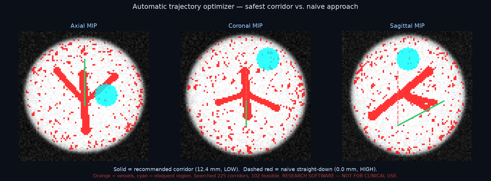
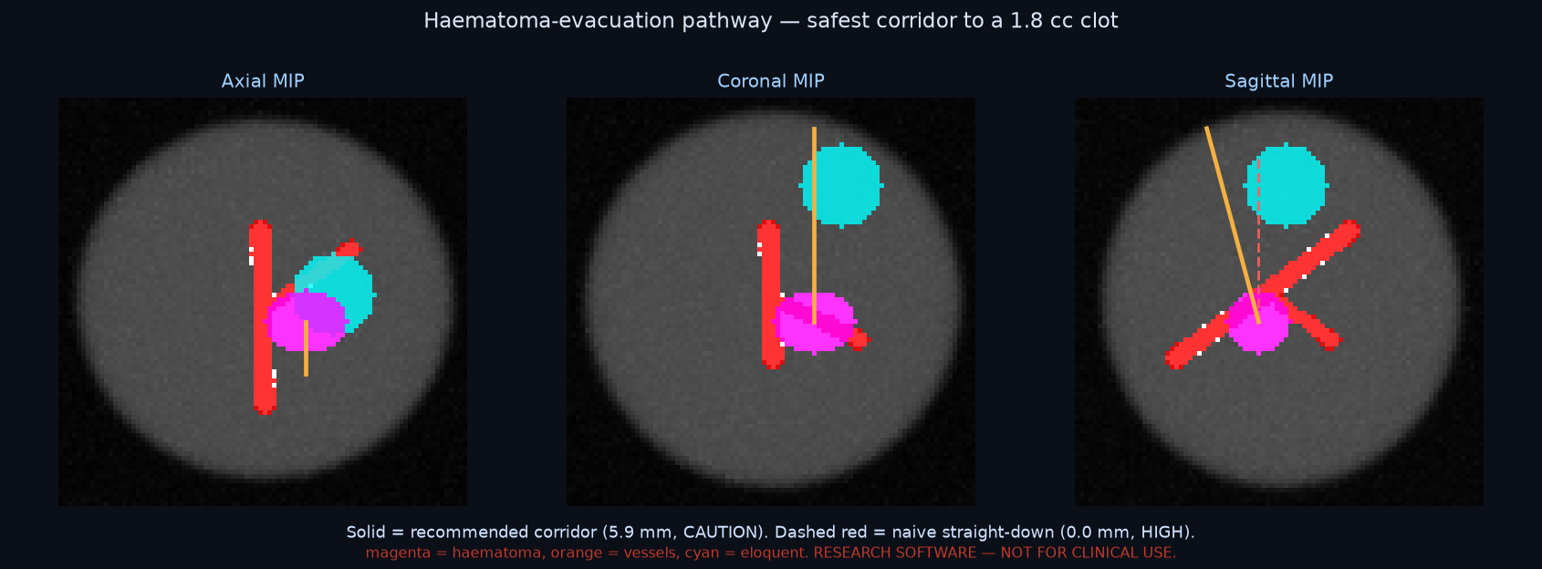
Name a deep target — including real DBS targets (subthalamic nucleus, globus pallidus) — and Sulcora scores candidate entry corridors against clearance to vessels, eloquent cortex, and tracts at once — optionally against predicted deformation — recommending the safest path with ranked alternatives, under per-structure margin, angle, and depth limits.
🔭
Probe-aligned views
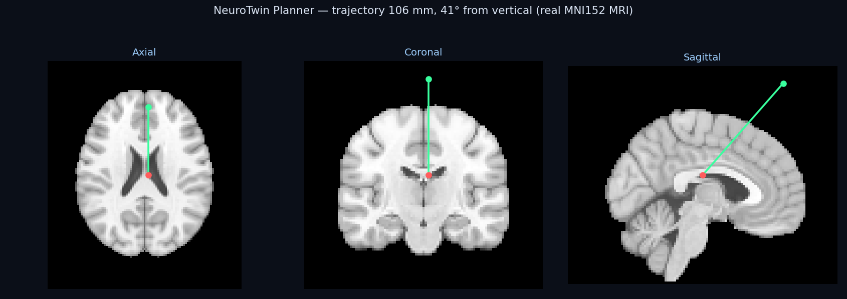
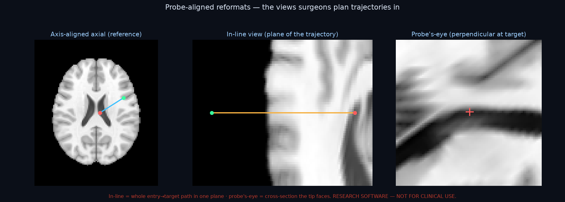
Beyond axial/coronal/sagittal — the in-line view (the whole entry→target path in one plane) and the probe's-eye view (the cross-section the advancing tip faces): the reformats surgeons actually plan trajectories in.
🧠
Structure-aware risk
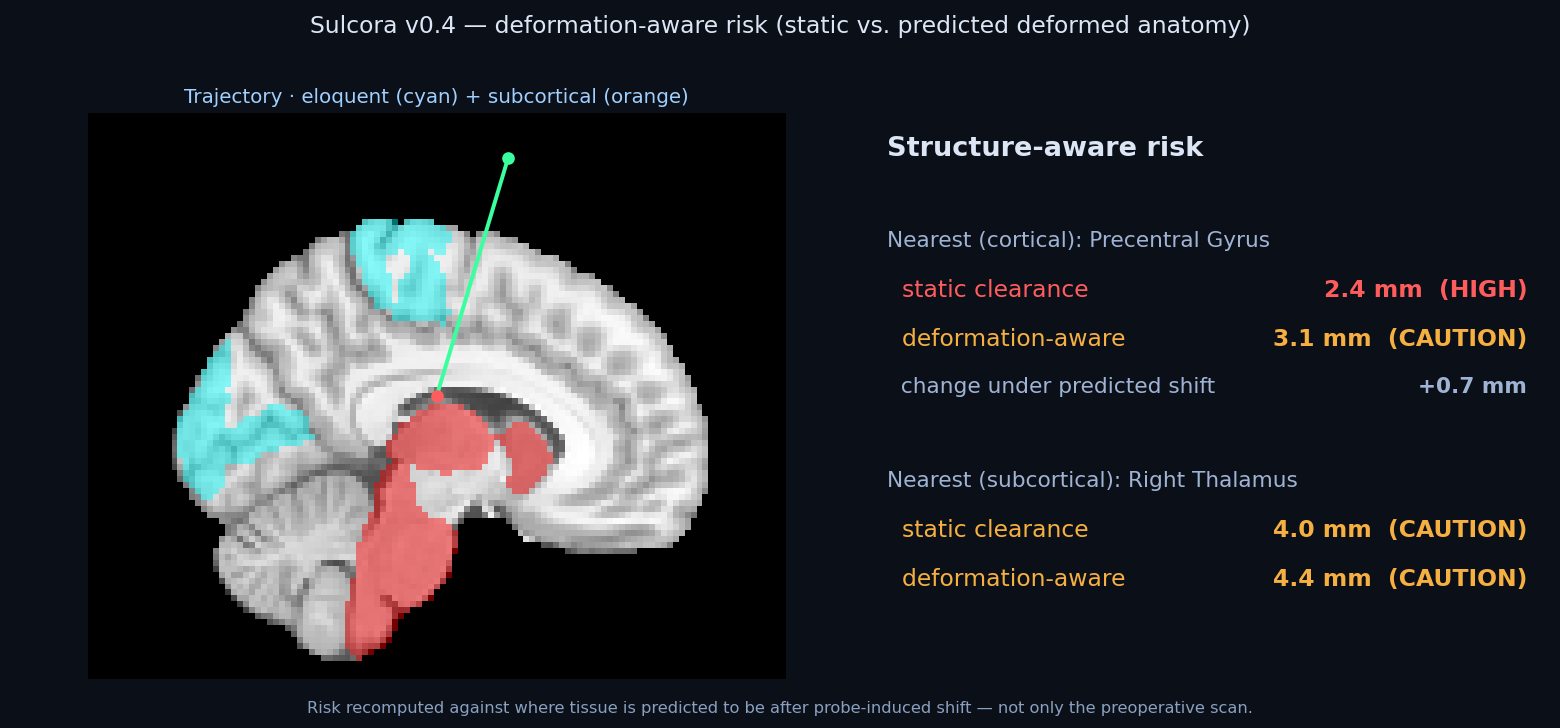
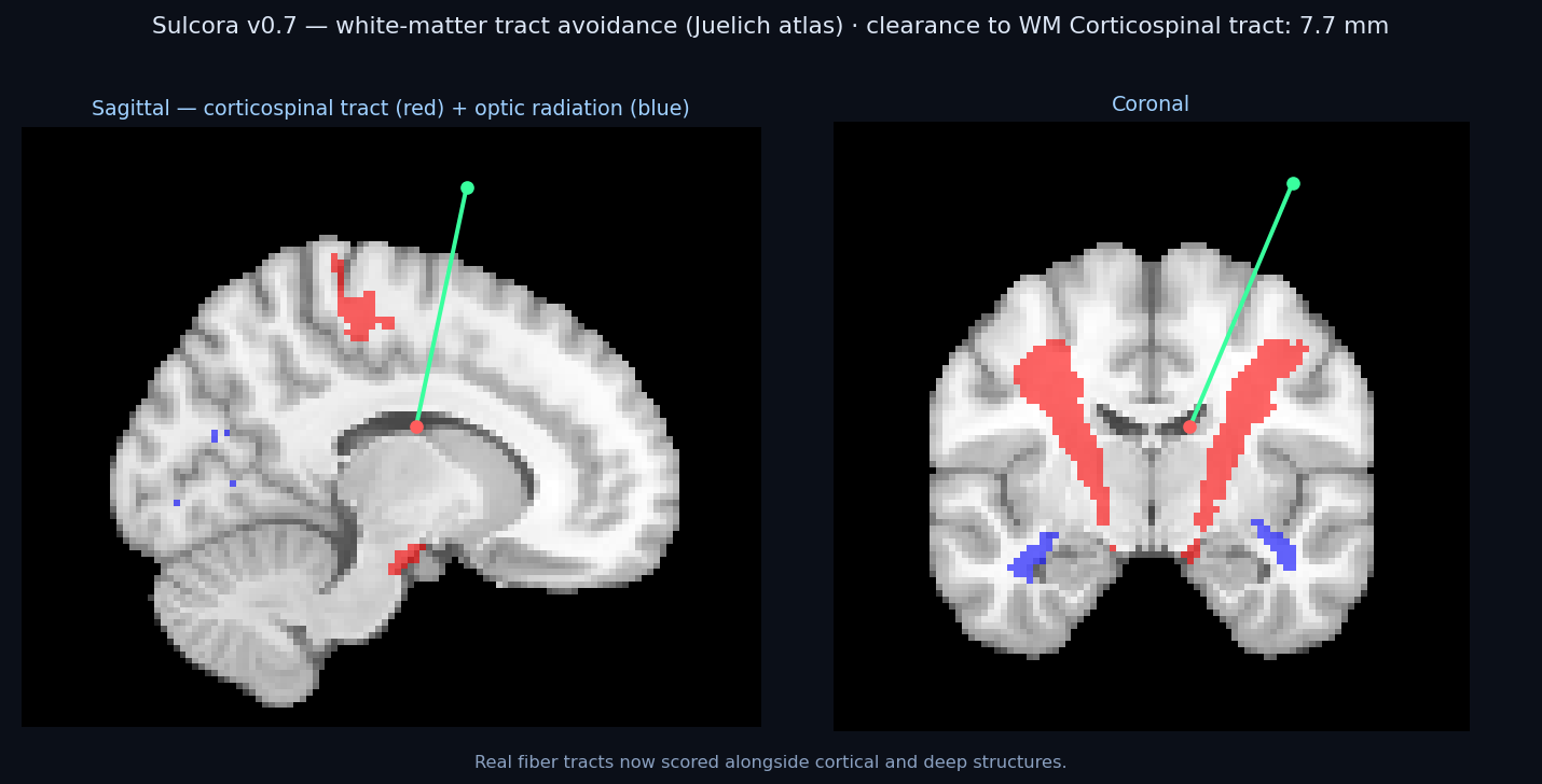
Real atlases: eloquent cortex (motor, speech, vision), deep structures (thalamus, hippocampus, brainstem), and white-matter tracts (corticospinal, optic radiation). Reports nearest structure, clearance, risk band.
🧩
Patient-specific segmentation
Optionally label eloquent cortex, ventricles, and deep structures directly from the patient's own T1 with a pretrained model (TotalSegmentator / SynthSeg) — registration-free, no atlas warp.
🩸
Vessel avoidance
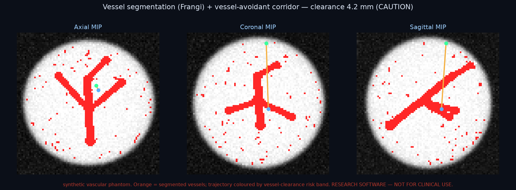
Segment the vasculature from CTA / MR-angiography (multiscale vesselness) and report clearance to the vessel tree — including against the predicted deformed anatomy. Hitting a vessel is the dominant depth-electrode and biopsy risk.
DIFFERENTIATORDeformation-aware risk
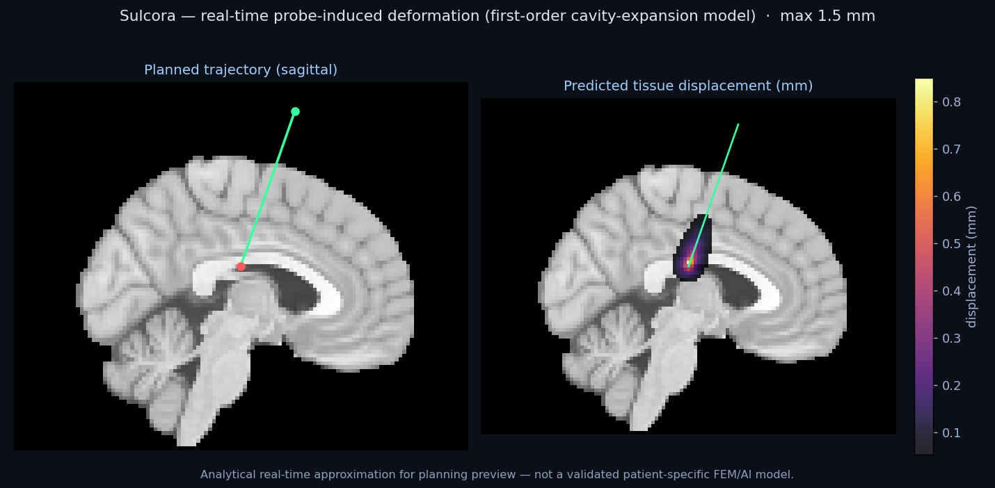
Risk recomputed against the brain's predicted deformed anatomy — the shift conventional static planning ignores.
🌊
Finite-element tissue deformation
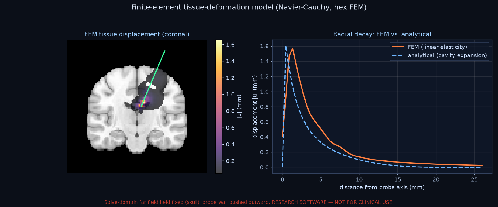
Predicts probe-induced tissue displacement by solving the elasticity equations on a finite-element mesh (Navier-Cauchy, hexahedral FEM) — upgrading the real-time analytical preview toward the validated model.
🎬
Insertion simulation
Advance the probe entry→target and watch clearance to critical structures at every depth, in all views.
ARPA-H TOPIC 7Robotics-validation bench
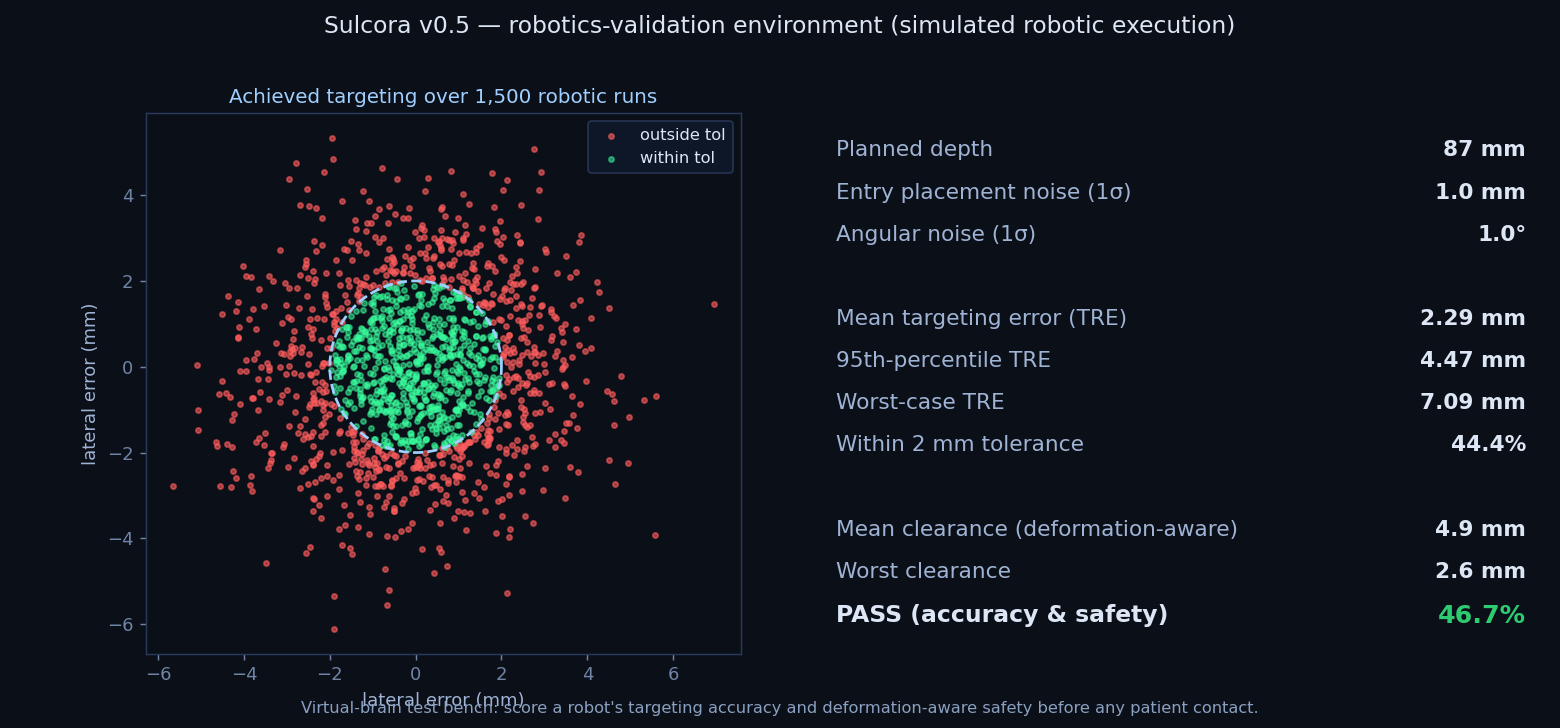
Score a simulated robot's targeting accuracy and deformation-aware safety over thousands of runs — before any patient contact.
⚿
Multi-trajectory planning
Plan multiple leads at once — bilateral DBS, multiple SEEG electrodes.
STROKE / ICHHaemorrhage (ICH) evacuation
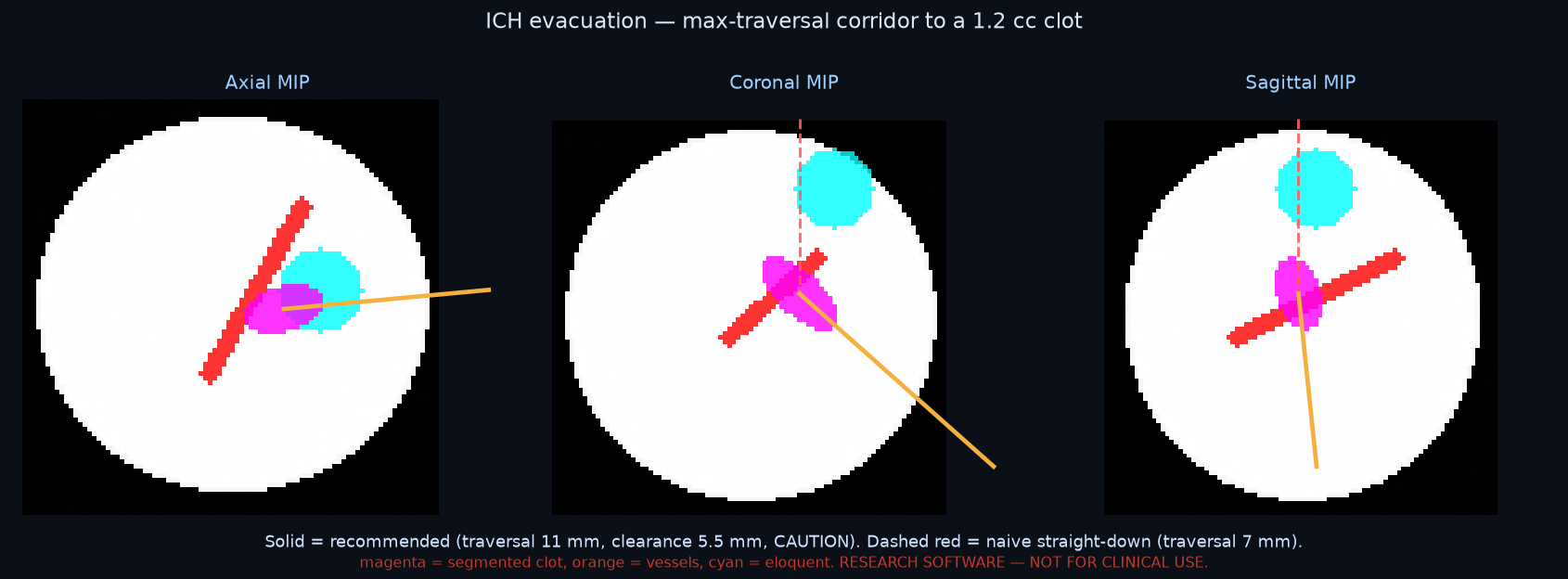
Segment an acute clot from CT, find its long axis, and plan the corridor that maximises clot traversal while clearing vessels and eloquent cortex — the minimally invasive approach shown to improve outcomes in the ENRICH trial (NEJM 2024).
🩹
Lesion procedures
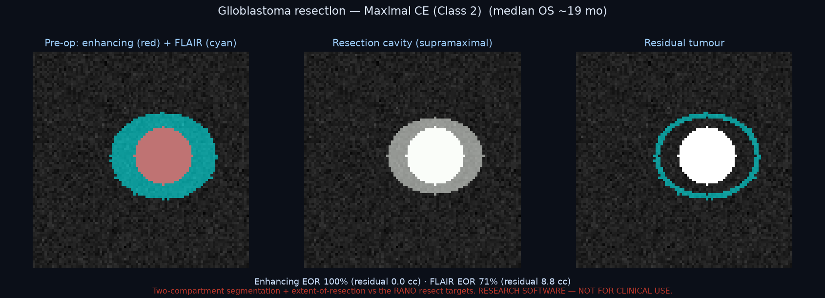
Beyond DBS/SEEG: target any segmented lesion by its centroid (volume in cc) for the safest corridor — biopsy, catheter/laser placement, or tumour access.
📄
Rich OR plan report
One-click PDF for the chart: per trajectory — tri-planar and probe-aligned reformats, a clearance-vs-depth profile, full geometry, and a per-structure clearance table (static and deformation-aware).
📏
Validation & interop
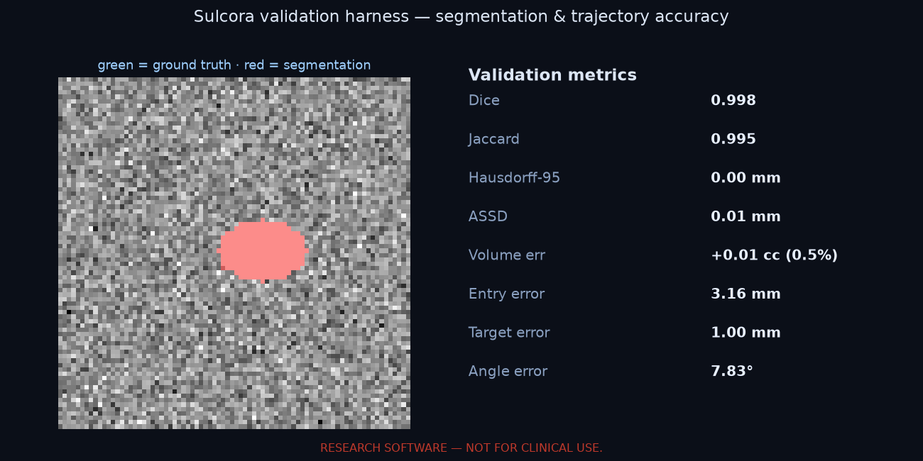
Quantitative accuracy metrics (Dice, Hausdorff-95, ASSD, volume & trajectory error) for evidence before clinical validation, and one-click export of the plan to 3D Slicer and a DICOM RT Structure Set for Brainlab / StealthStation.
🩻
Full CT support
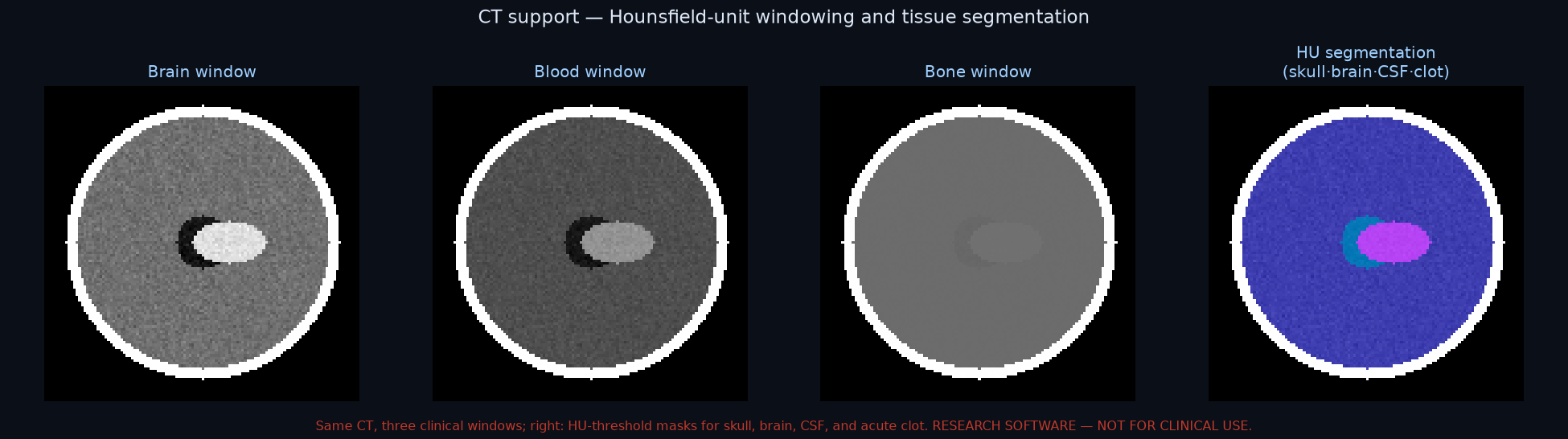
Head CT is the modality for haemorrhage and trauma — Sulcora loads it in Hounsfield Units with clinical window presets (brain / blood / bone / stroke) and HU-threshold tissue segmentation (skull, brain, CSF/ventricles, acute clot).
🔒
Local & private
Runs on the surgeon's own machine — DICOM (from PACS) or NIfTI opened from disk. The patient image is processed on-device and never leaves it: no cloud upload, no PHI in transit.